Have you already done a pediatric hip ultrasound examination?
D
evelopmental dysplasia of the hip (DDH) is one of the most common congenital deformities of the human locomotor system. It occurs due to abnormal growth and improper anatomical relationships of the joint bodies – the acetabulum on the pelvic bone and the head of the femur.
When we talk about developmental hip dysplasia, we refer to various forms of the disorder – from transient instability of the newborn hip, to poorly developed acetabulum (acetabular dysplasia), to partial (subluxation) or complete ( da se ne ponavlja) dislocation of the joint.
Risk factors
The exact cause of developmental hip dysplasia is unknown, but there are certain risk factors:
Presence of developmental hip dysplasia in close family members
More common in female children
Breech position in the womb or breech birth
Prematurity
Low amniotic fluid
Twin pregnancy
High birth weight
A higher incidence of developmental hip dysplasia is also seen in newborns with other musculoskeletal disorders, such as torticollis (congenital wry neck) or foot deformities (pes equinovarus, pes metatarsus adductus, etc.).

Ultrasound of children's hips
Early detection of developmental hip dysplasia is extremely important because it allows timely and successful treatment, preventing late consequences such as chronic pain, reduced range of motion in the hip joint, shortening of the affected leg, and limping.
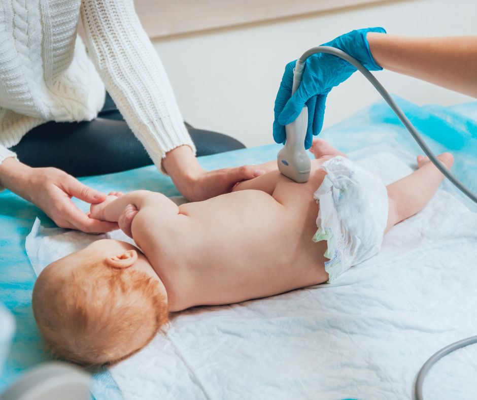
Therefore, in addition to regular clinical hip examinations in the maternity ward and during regular pediatric check-ups, it is recommended to routinely perform an ultrasound examination of the hips using the Graf method. This method provides a detailed insight into all anatomical structures of the joint and their relationships and is now an indispensable method in diagnosing developmental hip dysplasia.
The examination is recommended to be performed between the fourth and eighth week of life.
Preparation for the Hip Ultrasound Examination
The ultrasound examination is quick, painless, and completely harmless, with no exposure to radiation. The examination is performed by applying an ultrasound probe coated with contact gel to one hip and then the other. It is recommended to feed the child before the examination to ensure the baby is as calm as possible during the procedure. No additional preparation is required for the ultrasound examination.
This screening is conducted by trained pediatric orthopedists, pediatricians, or radiologists, and after a diagnosis is made, further treatment and monitoring are carried out by pediatric orthopedists.
The choice of treatment method and the success of the treatment depend on the degree of developmental hip dysplasia and the age at which it is detected. Therefore, the recommended screening ultrasound of the hips and regular systematic examinations are of utmost importance for every newborn.